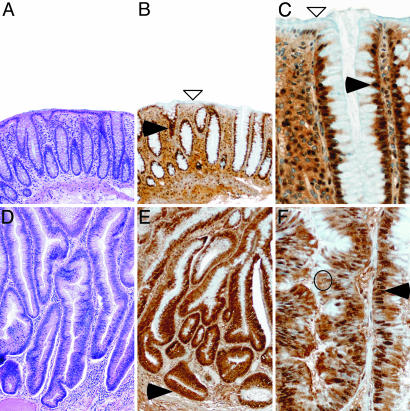
Fig. 4.
LRH-1 is differentially expressed in normal versus neoplastic colon. Shown is hematoxylin/eosin staining [A and D (magnification, ×80)] and LRH-1 immunostaining [B and E (magnification, ×80) and C and F (magnification, ×320)] of human normal colon mucosa (A–C) and neoplastic polyp (D-F). (A–C) In normal colon, LRH-1 is found predominantly in the nucleus of the crypt epithelial cells (filled arrowheads). LRH-1 immunostaining is detected in the epithelial cells lining the crypt compartment but absent in the surface epithelial cells (open arrowheads). (D–F) Neoplastic polyp characterized with high-grade dysplasia. A marked elongation and lack of differentiation and maturation of crypt and surface epithelial cells is observed. Nuclei are elongated and pseudostratified. Increased cytoplasmic (open circle) and nuclear (filled arrowheads) LRH-1 immunostaining is observed in all epithelial cells, including those lining the surface, which are normally nonproliferative and unstained.

