
Fig. 1. EC plasma membrane proteins are differentially expressed in OSA patients and OSA-free controls.
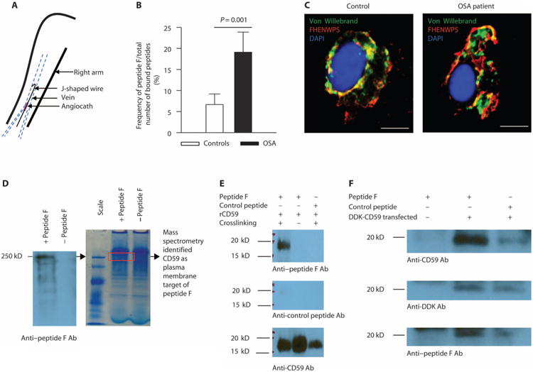
(A) Harvesting of venous ECs from study participants (n = 76 OSA patients and n = 52 controls, demographics in table S1). (B) Frequency of peptide F binding to ECs harvested from OSA patients [n = 18: 45% male; age, 44.6 ± 2.4 years; body mass index (BMI), 38.1 ± 2.1 kg/m2; apnea-hypopnea index (AHI), 26.2 ± 6.2 events/hour; oxygen desaturation index (ODI), 11.4 ± 2.8 events/hour] and controls (n = 20: 55% male; age, 40.9 ± 2.7 years; BMI, 27.9 ± 1.3 kg/m2; AHI, 1.3 ± 0.4; ODI, 0.37 ± 0.3 events/hour) expressed as a percentage of the total number of bound peptides (mean±SE; two-sided Student's t test adjusted for age, gender, and BMI shown in fig. S2, P = 0.001). (C) Representative confocal image of peptide F binding to ECs harvested from OSA patients (n = 10: 30% male; age, 50.3 ± 3.2 years; BMI, 34.9 ± 1.8 kg/m2; AHI, 19.4 ± 5.8 events/hour; ODI, 12.3 ± 3.3 events/hour) and controls (n = 10: 60% male; age, 38.4 ± 3.4 years; BMI, 29.4 ± 1.6 kg/m2; AHI, 1.9 ± 0.7 events/hour; ODI, 1.1 ± 0.6 events/hour); ECs are identified by immunofluorescence for von Willebrand factor. Scale bars, 10 μm. (D) Native polyacrylamide gel electrophoresis (PAGE) and Western blotting with anti–peptide F antibodies (Ab) of human umbilical vein endothelial cell (HUVEC) lysate incubated with or without peptide F. The region of bound peptide F (a single major band at molecular weight of 250 kD) was cut out from the gel (red rectangle), subjected to trypsin digestion, and analyzed by mass spectrometry. Among proteins identified in this region, CD59 was the only one that is expressed on the plasma membrane. (E) In vitro binding of re-combinant CD59 (rCD59; molecular weight, 18 to 20 kD) to peptide F (molecular weight, 700 to 800 daltons) after cross-linking but not to control peptide (Western blots probed with anti–peptide F, anti–control peptide, and anti-CD59 antibodies; n = 3). (F) Immunoprecipitation with beads coated with anti-DDK antibodies of human embryonic kidney (HEK) 293 cells transfected with Myc-DDK–tagged human CD59 plasmid incubated with peptide F, followed by SDS-PAGE and Western blotting with anti-CD59, anti-DDK, and anti–peptide F antibodies, showing specific binding of peptide F to CD59 (n = 3).