
Fig. 2. Cellular distribution of CD59 is altered in OSA.
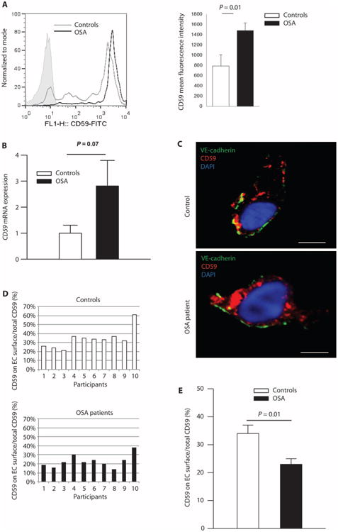
(A) Mean fluorescence intensity of total cellular CD59 (flow cytometry, shaded area represents isotype control; the quantity of cells on the y axis is expressed as percentage of total specimens). Bar graph quantitates total CD59 fluorescence (n = 8 OSA patients: 62% male; age, 42.3 ± 5.2 years; BMI, 35.9 ± 2.0 kg/m2; AHI, 21.3 ± 8.7 events/hour; ODI, 12.9 ± 4.5 events/hour; n = 9 controls: 33% male; age, 36.7 ± 4.6 years; BMI, 35.8 ± 3.2 kg/m2; AHI, 3.2 ± 0.7 events/hour; ODI, 2.0 ± 0.5 events/hour) (mean ± SE; two-sided exact permutation test). (B) Quantitation of the CD59 mRNA expression in ECs harvested from OSA patients (n = 21: 67% male; age, 45.5 ± 2.7 years; BMI, 35.6 ± 2.1 kg/m2; AHI, 23.5 ± 5.5 events/hour; ODI, 19.3 ± 6.1 events/hour) expressed as a fold change over controls (n = 15: 27% male; age, 34.1 ± 3.4 years; BMI, 36.6 ± 3.0 kg/m2; AHI, 1.3 ± 0.4 events/hour; ODI, 0.3 ± 0.2 events/hour) (two-sided Student's t test adjusted for age, gender, and BMI, shown in fig. S2). (C) Representative confocal images of cellular distribution of CD59 in ECs harvested from OSA patients (n = 10: 30% male; age, 50.3 ± 3.2 years; BMI, 34.9 ± 1.8 kg/m2; AHI, 19.4 ± 5.8 events/hour; ODI, 12.3 ± 3.3 events/hour) and controls (n = 10: 40% male; age, 34.5 ± 3.7 years; BMI, 34.1 ± 3.0 kg/m2; AHI, 2.2 ± 0.6 events/hour; ODI, 1.4 ± 0.6 events/hour); EC plasma membrane is identified by immunofluorescence for vascular endothelial (VE)–cadherin (CD144). Scale bars, 10 μm. (D) Histogram representing the percentage of total endothelial CD59 located on the plasma membrane for individual patients [n = 10 OSA patients; n = 10 controls; demographics in the legend for (C)]. (E) Quantitation of the percentage of total endothelial CD59 located on the plasma membrane [n = 10 OSA patients; n = 10 controls; demographics in the legend for (C)]. Linear regression confirmed that the difference in plasma membrane expression of CD59 is not confounded by age, gender, or BMI (fig. S2) (mean ± SE; two-sided exact permutation test). FITC, fluorescein isothiocyanate.