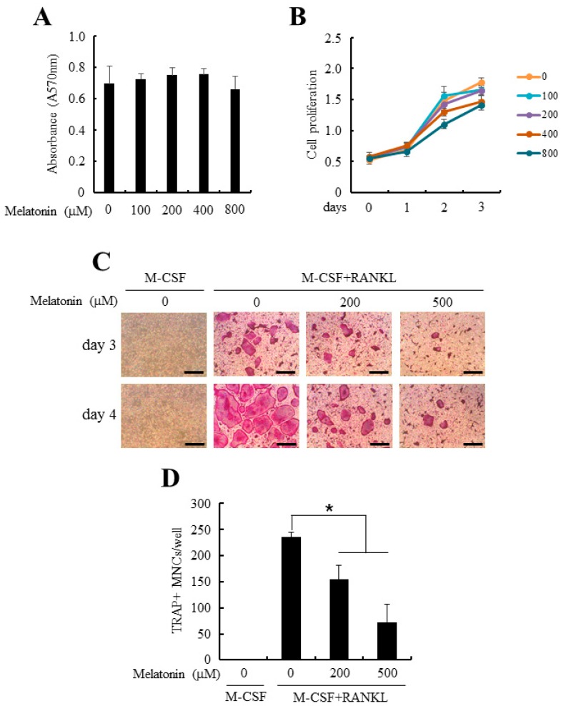
Figure 1.
Melatonin suppressed osteoclast differentiation from bone marrow-derived macrophages (BMMs). (A) Mouse BMMs were cultured in the presence of indicated doses of melatonin (0–800 µM). After 24 h of culture, cell viability was measured by MTT assay as described in Section 4; (B) BMMs were cultured in the osteoclastogenic medium (30 ng/mL macrophage colony-stimulating factor (M-CSF) + 100 ng/mL receptor activator of nuclear factor κB ligand (RANKL)) together with various concentrations of melatonin (0–800 µM) for 3 days. Cell proliferation was evaluated by 3-(4,5-dimethylthiazol)-2,5-diphenyltetrazolium bromide (MTT) assay; (C) BMMs were differentiated into osteoclast with osteoclastogenic medium in combination with or without melatonin (0–500 µM) for 4 days. At the end of culture, osteoclasts were stained for tartrate-resistant acid phosphatase (TRAP) activity. Bars, 200 µm; (D) TRAP+ multinucleated cells (TRAP+ MNCs) were quantified from experiments in panel (C). All quantitative data are presented mean ± standard deviation (SD), * p < 0.01).

