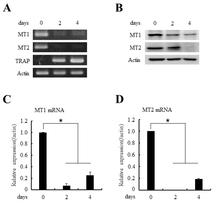
Figure 2.
The expressions of MT1 and MT2 melatonin receptors decreased during osteoclast differentiation. (A) BMMs were cultured with M-CSF (30 ng/mL) and RANKL (100 ng/mL) for indicated days. After the culture, total RNAs were isolated and the expressions of MT1/MT2 mRNAs were examined by RT-PCR (reverse transcription-polymerase chain reaction) analyses. TRAP served as a marker for osteoclast differentiation; (B) BMMs were cultured as in panel (A) and whole cell lysates were prepared and MT1/MT2 protein expressions were determined by immunoblotting with anti-MT1 or anti-MT1 antibodies; (C,D) The mRNA expressions of MT1 and MT2 were analyzed by quantitative real-time PCR after culturing BMMs in the 30 ng/mL M-CSF and 100 ng/mL RANKL for the indicated days. All quantitative data are presented mean ± SD (* p < 0.01).

