Figure 1.
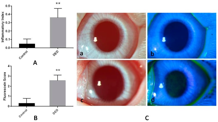
In vivo observations of ocular surface inflammation and corneal fluorescein staining in control and dry eye disease (DED) eyes. (A) The inflammatory index in the two groups. The DED group had a significantly higher inflammatory index than the control group; (B) the corneal fluorescein staining scores in the two groups. The DED group had a significantly higher fluorescein staining score than the control group; (C) photographs of corneas stained with and without fluorescein sodium. Signs of ocular abnormalities were not observed in the control eyes (a,b). Corneal edema and diffuse epithelial disruption were observed in the DED eyes (c,d) (16×). ** p < 0.01.

