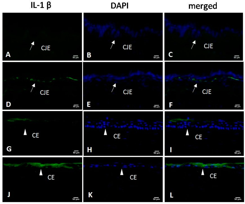
Figure 4.
Immunofluorescence staining with interleukin-1β (IL-1β)-specific antibodies in the conjunctival epithelium (CJE) and corneal epithelium (CE) (IL-1β staining: green; nuclear staining: blue). In the control group, IL-1β staining was very weak in the CJE (A–C) and localized only to the surface of the CE (G–I). CJE and CE were thinner in the DED group, in which the basal layer of the CJE (D–F) and the entire CE (J–L) stained positive for IL-1β (×400). CJE: conjunctival epithelium (white arrow); CE: corneal epithelium (white triangle). Scale bar = 50 μm.

