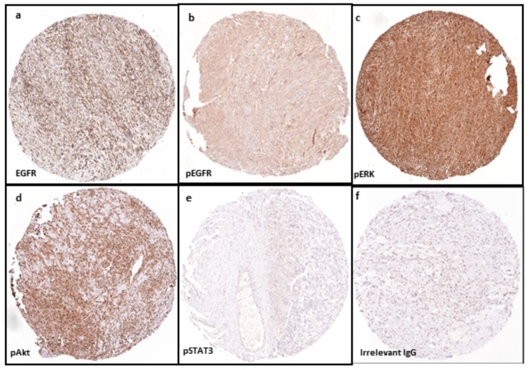
Figure 1.
Expression of protein markers using tissue microarray technology and immunohistochemistry in a sample of leiomyosarcoma (LMS) cores (amplification ×100): (a) positive expression (brown colour) of total EGFR; (b) phosphorylated EGFR; (c) pERK; and (d) pAkt in tissue array of LMS specimen. In contrast, negative immunohistostaining (blue colour) is shown in detection of: (e) pSTAT3 in LMS; and (f) control sarcoma sample (irrelevant IgG). Abbreviations: EGFR, epidermal growth factor receptor; pEGFR, phosphorylated EGFR; pERK, phosphorylated extracellular signal-regulated kinase; p-Akt, phosphorylated protein kinase B; pSTAT3, phosphorylated signal transducers and activators of transcription-3.

