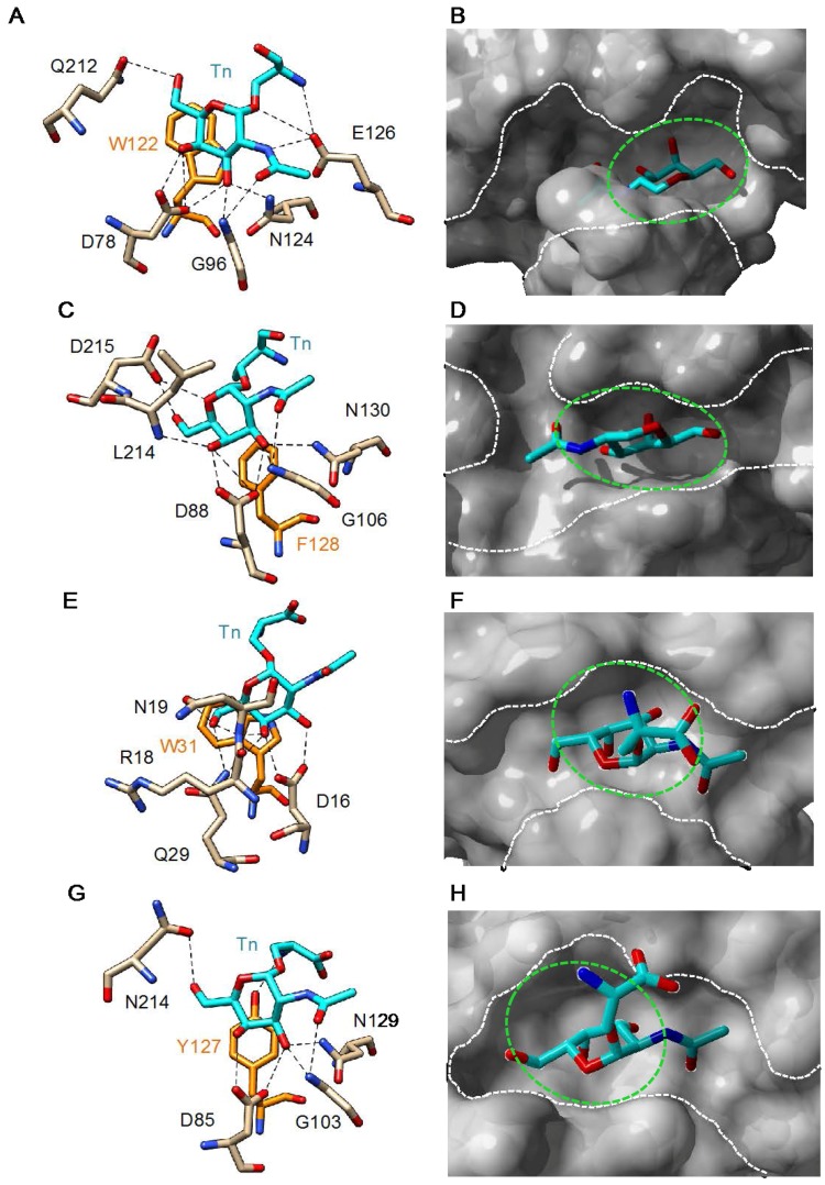
Figure 3.
(A,C,E,G) Network of hydrogen bonds and stacking interactions anchoring Tn antigen (Tn) to the monosaccharide-binding site of: Bauhinia forficata BfL lectin (A) (PDB code 5T5J) [46]; soybean lectin SBA (C) (PDB code 4D69) [89]; Sambucus nigra SNA-II lectin (E) (PDB code 3CA6) [74]; and Vicia villosa VVA-B4 lectin (G) (PDB code 1N47) [90]. Amino acid residues involved in stacking interactions with the disaccharide are colored orange; (B,D,F,H) Docking of Tn antigen to the monosaccharide-binding cavity (green dashed circle) of: Bauhinia forficata BfL lectin (B); soybean lectin SBA (D); Sambucus nigra SNA-II lectin (F); and Vicia villosa VVA-B4 lectin (H). The white dashed lines delineate the extended binding sites at the molecular surface of the different lectins. Cartoons drawn with Chimera [91].

