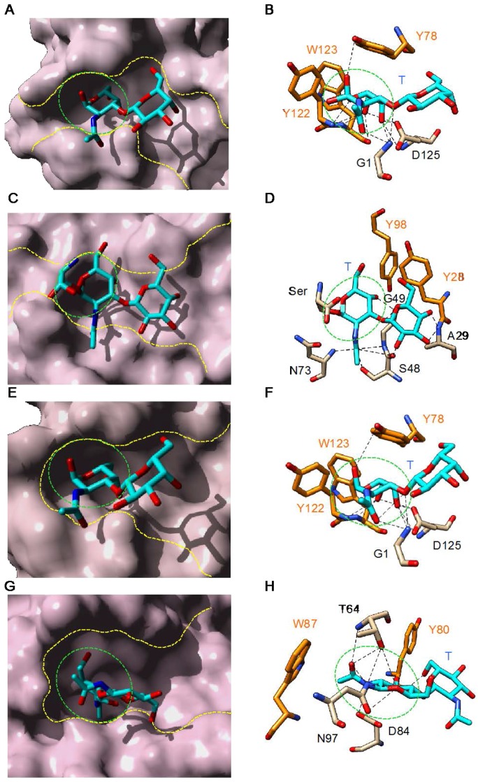
Figure 4.
Monosaccharide-binding sites (green dashed circles) and extended binding sites (yellow dashed lines) of: jacalin (Artocarpus integrifolia) (PDB code 1M26) [92] (A); the mushroom Agaricus bisporus lectin ABL (PDB code 1Y2V) [34] (C); the Osage orange (Maclura pomifera) lectin MPA (PDB code 1JOT) [58] (E); and the bitter gourd (Momordica charantia) galactose-specific lectin BGSL (PDB code 4ZGR) [62] (G), in complex with T-antigen (Galβ1→3GalNAcα1→Ser/Thr). Network of hydrogen bonds (dashed lines) anchoring T-antigen (colored cyan) to the amino acid residues of the extended binding site of: jacalin (B); ABL (D); MPA (F); and BGSL (H). Amino acid residues involved in non-polar stacking interactions with the disaccharide are colored orange. Cartoons drawn with Chimera [91].

