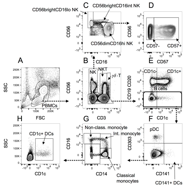
Figure 4.
The gating strategy applied to PBMCs analysed for monocytes, NK cells and DCs. (A) PBMCs were selected according to the forward scatter (FSC)/side scatter (SSC) profile; (B) NK cells, NKT and γδ-T cells were separated from CD56− cells, and from one another according to CD3 staining; (C) NK cells gated in B were separated into CD56brightCD16lo, CD56brightCD16int and CD56dim cells, according to CD16 and CD56 staining; (D) CD56dim NK cells were further separated into CD57+ or CD57− according to CD57 expression; (E) B cells were separated from non-B cells in the non-NK, NKT or γδ-T cell CD56- category identified in B. B cells were also divided into CD1c+ or CD1c− populations; (F) Non-B cells identified in E were divided into CD141+ myeloid DCs (mDC2), CD303+ plasmacytoid DCs (pDCs), or other cells. (G) Cells negative for CD141 and CD303 staining in F were separated based on CD16 and CD14 expression into classical (CD14hiCD16−), intermediate (CD14+CD16+) and non-classical (CD14loCD16+) monocytes (reference [23]), as well as CD14−CD16− cell types; (H) The CD14−CD16− cells identified in G were gated on CD1c+ to identify CD1c+myeloid DCs (mDC1).

