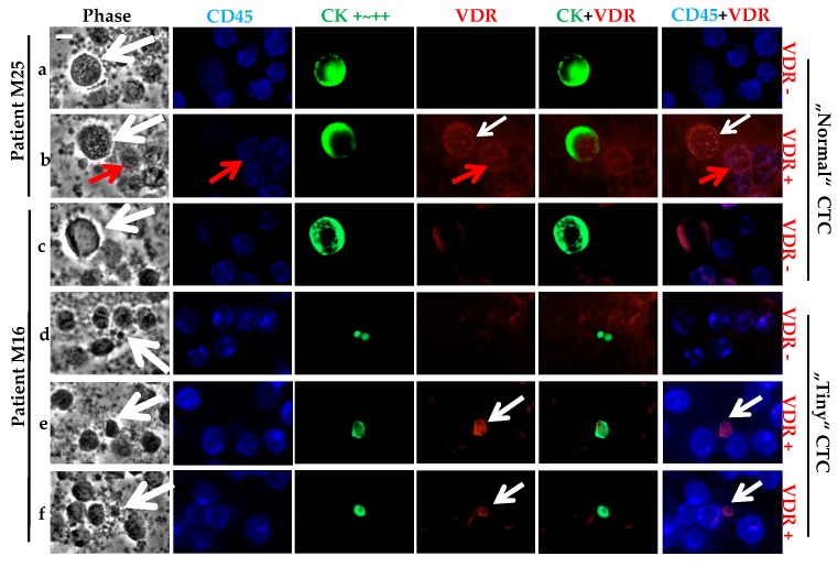
Figure 4.
VDR status determination on CTCs of metastatic BC patients. Triple fluorescence labeling of CD45 (in blue), CK (in green), and VDR (in red) was performed on 106 PBMCs, with parallel phase analysis. CTCs (with white arrows) were classified as VDR+ or VDR-. For both patients M25 (a,b) or M16 (c–f), either status was observed with superimposed VDR and CK labeling. CTCs exhibit size heterogeneity for patient M16 (“Normal” or “Tiny” CTCs). VDR staining was also seen on PBMCs (with red arrows), with superimposed VDR and CD45 labeling. Original magnification, ×40. Scale bar (white bar in the upper left image), 10 μm.

