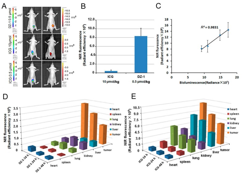
Figure 2.
The distribution of DZ-1 and ICG in the organs of subcutaneous tumor model. (A) Dual ex vivo BLI/NIRF imaging of mice with Hep3B-Luc subcutaneous xenografts treated with varying doses of ICG or DZ-1; (B) Quantification of NIRF intensity within the tumor area (per cm2) of subcutaneous tumor xenografts; (C) NIRF/BLI signal intensity correlation in mice (n = 5) with subcutaneous tumor xenografts (right); (D,E) Distribution intensity per cm2 of DZ-1 and ICG in the organs of subcutaneous tumor models at successive times.

