
Figure 3.
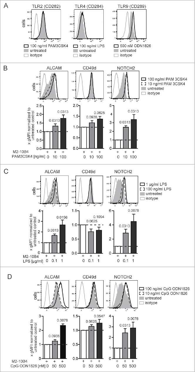
Activation of TLR-dependent signaling alters cell surface expression of ALCAM, CD49d, and NOTCH2 on Eμ-Tcl1 tumor cells. (A) 4 ×10⁵ Eµ-Tcl1 cells/mL were co-cultured with M2-10B4 cells and supplemented with either 100 ng/mL TLR1/2 ligand PAM3CSK4, 100 ng/mL TLR4 activator LPS, 500 nM TLR9 ligand ODN1826 or without. Surface expression of TLR2 (CD282), 4 (CD284), and intracellular expression of TLR9 (CD289) was assessed after 24 h. Representative histograms from n = 7 Eµ-Tcl1 leukemia cell clones analyzed in three independent co-culture experiments are depicted. (B–D) 4 × 10⁵ tumor cells/ml were supplemented with either (B) TLR1/2 agonist PAM 3CSK4, (C) LPS, or (D) stimulatory class B CpG ODN1826, a TLR9 agonist. Surface expression of ALCAM, CD49d, and NOTCH2 was assessed after 24 h. Per treatment 3–5 independent experiments were conducted with in total 6–10 tumor cell clones. Representative histograms of one clone are shown. Clone specific differences were normalized by dividing the gMFI of each treated sample with the gMFI of the corresponding untreated sample and bar diagrams represent means and SEMs of fold gMFIs. p values were calculated with the Wilcoxon signed rank test against a theoretical median of 1.