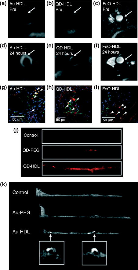
FIGURE 2.

Multimodality imaging of atherosclerosis using nanocrystals encapsulated in high density lipoprotein. T1-weighted MR images of the aorta of apoE KO mice pre- (a and b) and 24 h postinjection (d and e) with Au-HDL or QD-HDL. Arrows indicate areas enhanced in the post images. (c and f) T2*-weighted images of an apoE KO mouse pre- and 24 h postinjection with FeO-HDL. (g–i) Confocal microscopy images of aortic sections of mice injected with nanocrystal HDL. Red is nanocrystal HDL, macrophages are green, and nuclei are blue. Yellow indicates colocalization of nanocrystal HDL with macrophages and is indicated by arrowheads. (j) Fluorescence image of aortas of mice injected with QD-HDL, QDPEG, and saline. (k) Ex vivo sagittal CT images of the aortas of mice injected with Au-HDL, Au-PEG, and saline. (Reprinted with permission from Ref 95. Copyright 2008 American Chemical Society)
