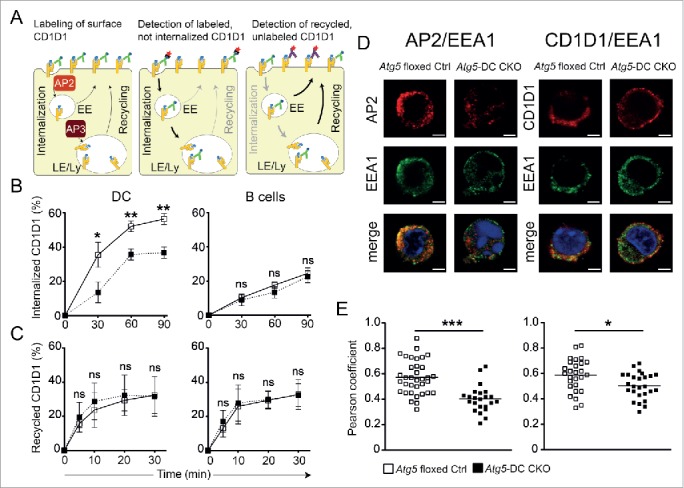
Figure 4.

Impaired CD1D1 internalization in Atg5-DC CKO DCs. CD1D1 surface levels are determined by the rates of CD1D1 internalization and recycling through endosomal and lysosomal compartments. EE, early endosomes; LE, late endosomes; Ly, lysosomes (A). Internalization of surface CD1D1 on splenic DCs or splenic B cells was analyzed using a biotin-based flow cytometric endocytosis assay. Pooled data and SEM of at least 3 independent experiments are shown. Each experiment contained at least 2 animals per group. Statistics: 2-tailed unpaired Student t test (B). CD1D1 recycling in splenic DCs or splenic B cells was analyzed using a flow cytometry based recycling assay. Pooled data and SEM of at least 3 independent experiments are shown. Each experiment contained at least 2 animals per group. Statistics: 2-tailed unpaired Student t test (C). Colocalization study of AP2 and EEA1, as well as CD1D1 and EEA1 via confocal microscopy. Original magnification with 63×, 1.4 NA oil immersion lens. Representative photographs from 2 independent experiments per colocalization study are shown. Scale bar: 2.5 µm (D). Scatter dot plot representation and quantification of colocalization between AP2 and EEA1 and between CD1D1 and EEA1 via the Pearson coefficient. Each symbol represents one cell. Pooled data of 2 independent experiments per colocalization study are shown. Statistics: 2-tailed unpaired Student t test (E). *** P≤ 0.001,** P≤ 0.01,* ≤ 0.05, ns P> 0.05.
