
Figure 3.
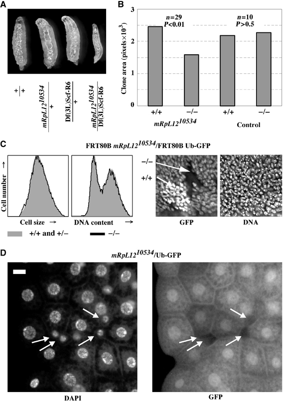
Homozygous mRpL1210534 cells have a cell-autonomous growth defect. (A) Images of larvae at 70 h AED. (B) Homozygous mRpL1210534 or control clones were induced at 66 h and dissected at 114 h AED, and wing discs were stained with DAPI and imaged. The area without GFP (−/−) and two copies of GFP (+/+) was measured in Photoshop. Genotypes: hs-Flp122; FRT80B mRpL1210534 or FRT80B/FRT80B Ub-GFP13A. (C) Wing discs from (B) were analyzed by FACS and GFP-negative cells (black line) were separated from GFP-positive cells (one or two copies; filled gray histogram. Shown are the forward scatters and DNA contents blotted against cell numbers (left). DAPI and GFP staining of a representative FRT80B mRpL1210534/FRT80B Ub-GFP13A twinspot (right). (D) Homozygous mRpL1210534 cells were induced in the fat body during embryogenesis by ionizing radiation, and third instar larvae were dissected and their fat body mounted. Homozygous mutant cells are marked by the absence of GFP (white arrows). Scale bar, 10 μm.