
Figure 5.
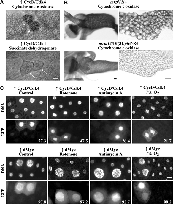
CycD/Cdk4 stimulates mitochondria in fat body cells. (A) COX (top) and SDH (bottom) activity in fat body cells. CycD/Cdk4-expressing cells were detected by coexpression of GFP in random clones, and are marked by arrows. (B) COX staining in second instar larvae, 77 h AED for the midgut and the proventriculus (left) and 96 h AED for the fat body (right). Images of heterozygous (mrpl1210534 or Df(3L)Scf-R6/+) or homozygous mutant (mrpl1210534/Df(3L)Scf-R6) larvae were exposed and treated identically in Photoshop. (C) Random clones expressing CycD/Cdk4 (top) or dMyc (bottom) were induced as described in Figure 2E, and marked by coexpression of GFP. At 24 h AED, larvae were transferred to normal food, or food supplemented with rotenone (5 μg/ml), antimycin A (5 μg/ml), or normal food and larvae were incubated at 7% O2. All were dissected at 116 h AED, fixed and stained with DAPI, and fat bodies were mounted. Numbers indicate the percentages of GFP-positive cells that are increased in ploidity, compared to neighboring cells. Quantifications were carried out in blind (n⩾150). Scale bar, 20 μm.