
Figure 4.
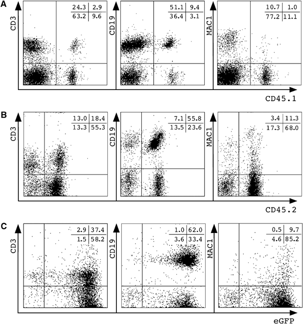
Lymphoid/myeloid donor cells in recipients after transplantation of bulk TSA/AzaC-treated neurosphere cells. Shown are splenocytes of wt (A) or eGFP/bcl2 (B) neurosphere transplant recipients stained with CD45 donor-type and lineage marker antibodies. (C) FACS analysis of the peripheral blood of a representative secondary CD45.1 recipient (n=3) receiving bone marrow cells of animal #154. Gated CD45.1− cells were plotted against lineage markers and eGFP.