
Figure 5.
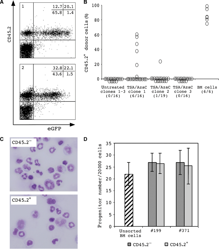
Donor cells in transplant recipients of cloned TSA/AzaC eGFP/bcl2 neurosphere cells. (A) FACS analysis of peripheral blood cells of two representative TSA/AzaC clone 1 recipients 3 months post-transplantation (panel 1: recipient #197; panel 2: recipient #199). (B) Percentage CD45.2+ donor cells in the peripheral blood 5–8 weeks post-transplant. Numbers of chimeric animals per total number of analysed animals are indicated. (C) Morphology of sorted recipient (CD45.2−) and neurosphere-derived (CD45.2+) cells from chimeric bone marrow of a clone 2 recipient. (D) Erythro/myeloid colony formation of sorted recipient (CD45.2−) and clone 1-derived (CD45.2+) bone marrow cells (transplant recipients #199 and #371).