
Figure 6.
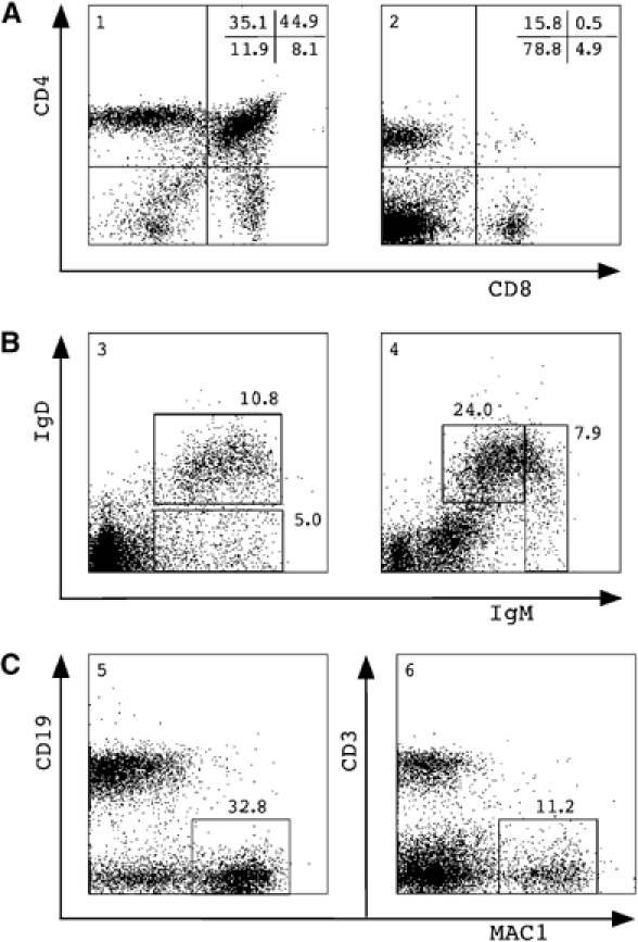
Lymphoid/myeloid repopulation by cloned TSA/AzaC eGFP/bcl2 neurosphere cells. (A) Thymocytes (panel 1) and splenocytes (panel 2) and (B) bone marrow cells (panel 3) and splenocytes (panel 4) were isolated and stained; gated eGFP+ donor-derived cells were stained with antibodies against CD4 and CD8 (A) or IgD and IgM (B). (C) Gated eGFP+ donor cells in bone marrow were stained with antibodies against CD19 and MAC1 (panel 5), and donor splenocytes were stained for CD3 and MAC1 (panel 6). Percentages of gated eGFP+ cells plotted against lineage-specific staining are shown. Analysis of a representative clone 1 recipient 3 months post-transplant is shown.