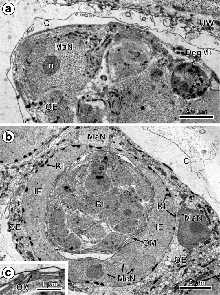
Fig. 2.
Initial stages of the outer and inner envelope formation. a Part of an early embryo adjacent to uterine wall. Note (1) already formed membraneous vitelline capsule surrounding an early embryo composed of several blastomeres of different sizes and (2) a large macromere, situated under the vitelline capsule at the periphery of other blastomeres, which contains predominant nucleus with spherical electron-dense nucleolus, which takes part in the outer envelope formation. b Preoncospheral phase in more advanced stage of embryonic development. Four primary embryonic envelopes (vitelline capsule, outer envelope, inner envelope and oncospheral membrane) are clearly visible. In the outer envelope, note large nucleus of macromeres which predominant nucleolus, numerous elongated mitochondria and higher concentration of free ribosomes. In the inner envelope, note three nuclei of mesomeres, the fusion of which forms the cytoplasm of the syncytial layer. c Detail of the oncospheral membrane. Bl blastomere, C vitelline capsule, DegMi degenerating micromere, IE inner envelope, KI keratin-like protein islands, MaN macromere nucleus, MeN mesomere nucleus, n nucleolus, OE outer envelope, OM oncospheral membrane, UW uterine wall

