
Figure 6.
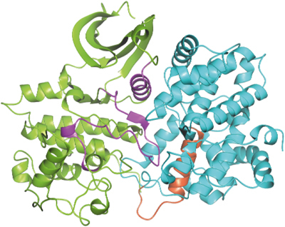
Structure of pCDK2 (in green) in complex with cyclin E1 (in cyan) with the 20-amino-acid sequence (residues 230–249) that are involved in centrosome localisation (Matsumoto and Maller, 2004) shown in red. The PSTAIRE (C-) helix and the activation segment, the two major regions of contact between pCDK2 and cyclin E1, are shown in magenta.