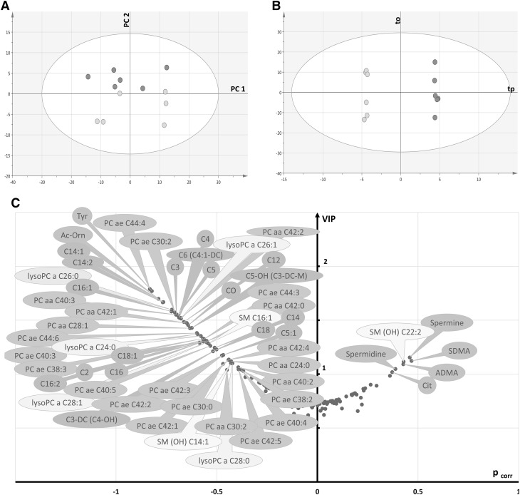
Fig. 4.
Multivariate analysis of control retinas: a first principal plan of the PCA (axis 1 vs. axis 2) showing no clear outlier and a separation between males (green circles) and females (pink circles), b OPLS-DA scatter plot showing a clear discrimination between the two groups along the predictive component tp. c Volcano plot of metabolites that best discriminate groups. They are essentially belonging to glycerophospholipids (dark and light orange bubbles) and acylcarnitines (brown bubbles), both relatively increased in male retinas, along with some sphingomyelins (yellow bubbles), amino-acids and biogenic amines (green bubbles). Legend: (in what follows, X indicates the length of the acyl chain and Y the degree of unsaturation) CX:Y acyl-l-carnitines, PC aa CX:Y phosphatidylcholine diacyl, PC ae CX:Y phosphatidylcholine acyl-alkyl, lysoPC a CX:Y lysophosphatidylcholine acyl, SM CX:Y sphingomyelin, SM(OH) CX:Y hydroxysphingomyelin, C0 carnitine, C2 acetyl-l-carnitine, C3 propionyl-l-carnitine, C3-DC (C4–OH) malonyl-l-carnitine (hydroxybutyryl-l-carnitine), C4 butyryl-l-carnitine, C5 valeryl-l-carnitine, C5:1 tiglyl-l-carnitine, C5–OH (C3-DC-M) hydroxyvaleryl-l-carnitine (methylmalonyl-l-carnitine), C6 (C4:1-DC) hexanoyl-l-carnitine, C12 dodecanoyl-l-carnitine, C14 tetradecanoyl-l-carnitine, C14:1 tetradecenoyl-l-carnitine, C14:2 tetradecadienyl-l-carnitine, C16 hexadecanoyl-l-carnitine, C16:1 hexadecenoyl-l-carnitine, C16:2 hexadecadienyl-l-carnitine, C18 octadecanoyl-l-carnitine, C18:1 octadecenoyl-l-carnitine, Ac-orn acetylornithine, ADMA asymmetric dimethylarginine, Cit citrulline, SDMA symmetric dimethylarginine, Tyr tyrosine

