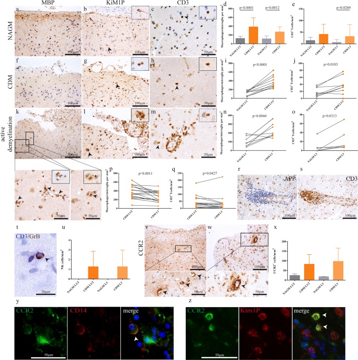
Fig. 1.
Increased adaptive and innate immune cell infiltration in demyelinated as compared to normal appearing cortical gray matter. In comparison with regularly myelinated normal appearing cortical gray matter (a–c), demyelinated cortex obtained at biopsy (f–h) shows higher densities of KiM1P+ macrophages/activated microglia (d, g, i, n) and T cells (e, h, j, o). An example of a cortical lesion with ongoing demyelination as evidenced by MBP-laden phagocytes (arrows, k–m). In addition to foamy macrophages (k, l), T cells are found perivascularly, as well as in the parenchyma (m). A gradient of macrophage/microglia (p) and T cell densities (q) is apparent with higher densities in the superficial cortical layers 1/2 compared to layer 3. Multilayered perivascular T cell cuffs in the cortex, identified by the presence of APP+ neurons, were only seen in rare cases (r, s). The patient shown here presented with neuropsychiatric symptoms (patient no. 25). GrB+/CD3− natural killer (NK) cells were observed perivascularly and only in demyelinated, but not normal appearing cortex (t, u, arrow). CCR2+ monocytes were mainly observed perivascularly, with some cells scattered in the parenchyma (v, w, x). CCR2/CD14 double-positive cells were observed perivascularly in patients with ongoing demyelination, indicating active myeloid cell recruitment into the cortex (y, arrows). Fluorescent double-immunohistochemistry of CCR2 and the pan-macrophage marker KiM1P identifies CCR2/KiM1P double-positive cells (z). All data are presented as mean ± SD. d, e One-way ANOVA followed by Dunn's Multiple Comparison post hoc test; i, j, n paired t test; o Wilcoxon signed-rank test; p, q paired t test and Wilcoxon matched pairs signed-rank test. All immunohistochemistries were performed with diaminobenzidine (DAB; brown color), except for CD3 (Fast Blue, in double labelling with GrB). Cell nuclei are counterstained with hematoxylin. APP amyloid precursor protein, NAGM normal appearing cortical gray matter, CDM cortical demyelination, GrB granzyme B, L1/2 cortical layer 1 and 2, L3 cortical layer 3

