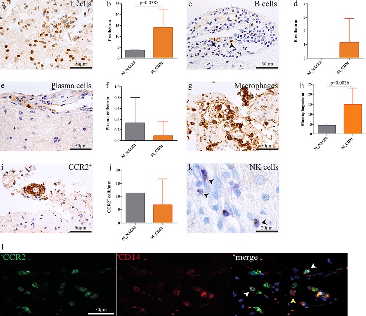
Fig. 2.
The intensity of meningeal inflammation associates with cortical demyelination. Meningeal inflammation, mostly arranged in a perivascular fashion, was found adjacent to demyelinated and non-demyelinated cortical areas. T lymphocyte (a, b) and macrophage (g, h) infiltration was significantly higher in areas overlying demyelinated cortex. Also, macrophages and T cells were more abundant than B cells (c, d) and plasma cells (e, f). NK cells were exceedingly rare (k). CCR2+ monocytes were found perivascularly in the leptomeninges overlying both demyelinated and normal-appearing cortical gray matter (i, j). Meningeal CD14+ CCR2+ and CD14+ CCR2− monocytes overlying an actively demyelinating cortical lesion in a patient with monocyte invasion into the subpial cortex (l). Data are presented as mean ± SD; Mann–Whitney test

