Fig. 6.
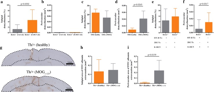
Encephalitogenic T cells are dispensable for subpial but not for perivascular demyelination. a, b Quantitative analysis of subpial and perivascular cortical demyelination in 8–18C5 (n = 8) and control antibody (n = 9) transferred RAG1−/− mice analyzed in two independent experiments. Data are presented as mean ± SD; unpaired t test. c, d Quantitative analysis of subpial and perivascular cortical demyelination in healthy (n = 4) and diseased OSE mice (n = 8) analyzed in two independent experiments. Data are given as mean ± SD; Mann–Whitney test. e, f Quantitative analysis of subpial and perivascular cortical demyelination in 2D2 Tc/8-18C5 antibody transferred RAG1−/− mice (n = 6) and OT-II Tc/8-18C5 antibody transferred RAG1−/− mice (n = 12). Representative data of two independent experiments are presented as mean ± SD; Mann–Whitney test. g Assessment of FITC-albumin extravasation by IHC in healthy (top) and diseased Th/+ mice (bottom) 24 h after stereotactic cytokine injection into the motor cortex. Dotted lines mark the cortical area where FITC-albumin extravasation was quantified. h, i Quantification of FITC-albumin extravasation in healthy (n = 4) and diseased Th/+ mice (n = 5). Data are presented as mean ± SD; Mann–Whitney test

