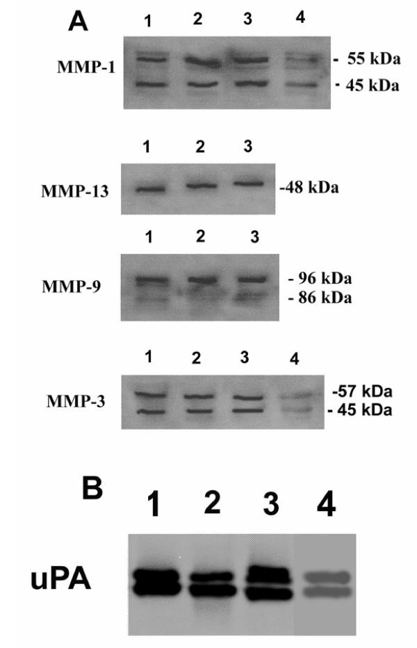
Figure 7.
Immunological characterization of MMPs and uPA in Breast Cancer Cells. Breast cancer cells (105 cells/well) stimulated with TGFβ (10-10 M) were cultured for 24 h in serum-free medium in the presence of 2 μg/ml of human plasminogen and CT1166 and aprotinin. Western blot analysis was undertaken as described in the Materials and methods section. Lane 1, MDA-231 cells; lane 2, ZR-75-1cells; lane 3 MCF-7 cells; lane 4, HME cells. Pro- and active forms of collagenase-1 gelatinase-B, and stromelysin-1 and proform of collagenase-3 were detected (A). Pro and active forms of uPA are shown (B).

