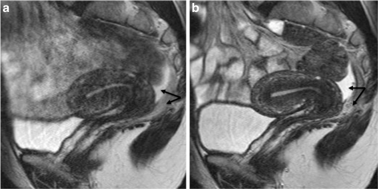
Fig. 2.
Sagittal 2D T2-weighted MR images performed at 1.5 Tesla showing the benefits of anti-peristaltic agents on image quality. Imaging performed in the same patient before (a) and after (b) administration of glucagon demonstrating a dramatic improvement in image quality. Note the presence of pelvic fluid in the pouch of Douglas underlining a clear demarcation between peritoneal and posterior subperitoneal compartments (double arrow) (reprinted with permission - Bazot M. Ed. Lavoisier-Paris 2016)

