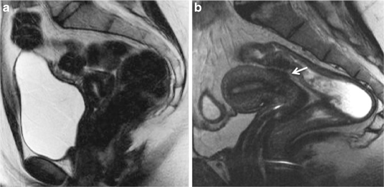
Fig. 3.
Sagittal 2D T2-weighted MR images performed at 1.5 Tesla showing the benefits of patient preparation on image quality. (a) Imaging performed with a full urinary bladder and without bowel preparation is sub-optimal for interpretation and disease may be overlooked. (b) MR imaging performed in a different patient following bowel preparation with Normacol and 2 h after emptying her urinary bladder. Note the superior image quality in (b) and the large endometriotic lesion on the anterior rectosigmoid colon (arrows)

