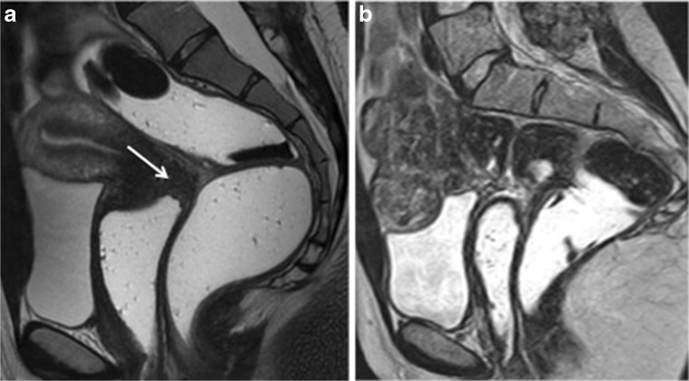
Fig. 4.
Sagittal 2D T2-weighted MR images performed in two different patients at 1.5 Tesla following vaginal and rectal opacification with sonographic gel and with (a) or without (b) bowel preparation. Vaginal distension demonstrates thickening of the posterior vaginal fornix (white arrow) without involvement of the pouch of Douglas or rectum posteriorly that is clearly analysable (a). Vaginal and rectal opacification without bowel preparation cannot permit an accurate analysis of potential deep posterior endometriosis, especially potential rectal endometriosis (b)

