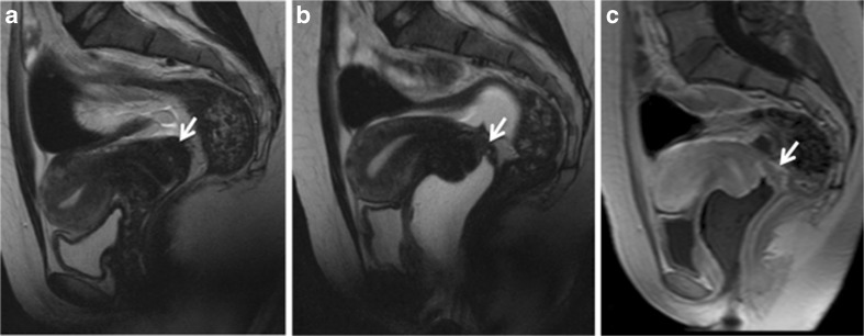
Fig. 5.
Sagittal 2D MR images performed at 1.5 Tesla demonstrating the use of sonographic gel to opacify and distend the vagina. (a) Sagittal 2D T2-weighted image demonstrating an endometriotic plaque involving the posterior vaginal fornix (white arrow). Following distension of the vagina with sonographic gel, the plaque is better delineated on both T2-weighted (b) and fat-suppressed T1-weighted (c) sequences (white arrows) (reprinted with permission - Bazot M. Ed. Lavoisier-Paris)

