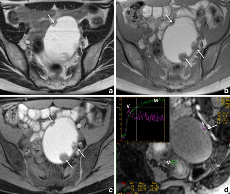
Fig. 6.
Axial 2D MR images performed at 1.5 Tesla demonstrating the use of gadolinium in the diagnosis of indeterminate adnexal mass related to endometrial cyst complicated with clear cell carcinoma. (a) Axial 2D T2-weighted image demonstrates a large unilocular cyst containing papillary projections and/or solid portion (arrows). Axial without (b) and with (c) fat-suppressed T1-weighted sequences display high signal content related to endometriotic fluid. Axial oblique dynamic contrast enhanced MR images (d) display location of region of interest (ROI) within external myometrium (M) and vegetation (V) and the initial increase in the signal intensity of solid tissue (arrow) that is steeper than that of myometrium (M), corresponding to a curve type 3 (V) highly suggestive of carcinoma confirmed at histopathological examination

