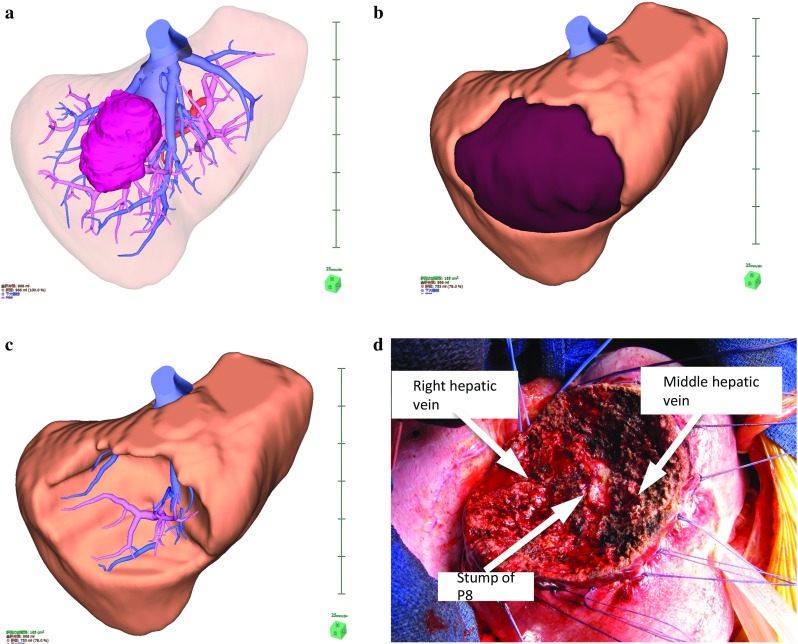
Fig. 2.

A case of segment 8 segmentectomy. A 3D image was generated from patient CT DICOM data using a 3D image analysis system. A large tumor located in segment 8 of the liver is shown (a). S8 segmentectomy was planned, and the resection line was drawn along the demarcation line of P8 (b). An image of the resected liver (c). The position of the stump of P8 and the running directions of the middle hepatic vein and the right hepatic vein were similar to those determined in the preoperative simulation (d)
