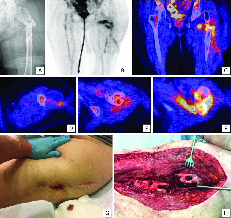
Fig. 3.
Clinical example of FDG-PET/CT. A 77-year-old woman who had a proximal femur fracture for which she underwent open reduction and internal fixation with a femur plate which had to be removed at a later stage due to infection. a X-ray, AP view: no consolidation, severe angulation, heterogeneous sclerotic aspect around the fracture. She was referred to our hospital with a fistula in the lateral thigh and a clinical suspicion of osteomyelitis of the proximal femur. Further imaging demonstrated an infection of the proximal femur, a medial abscess and a fistula coursing to the lateral aspect of the thigh which correlated with the clinical findings during surgery. b–f 18F FDG-PET/CT (b coronal FDG-PET image, c coronal fused FDG-PET/CT image, d–f transaxial fused FDG-PET/CT images). g clinical pre-operative picture, h perioperative clinical picture

