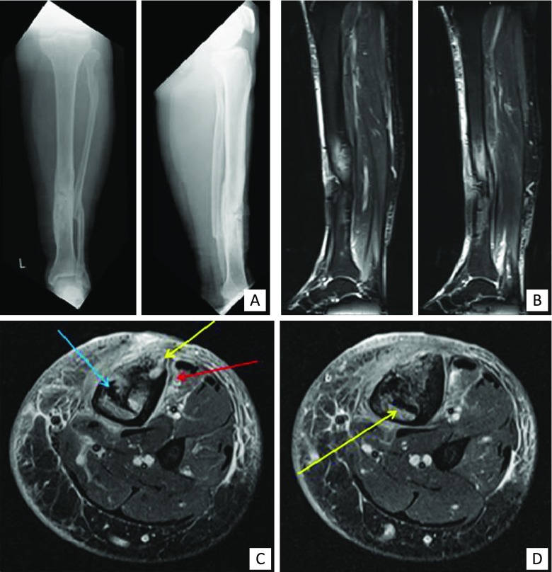
Fig. 4.
Clinical example of MRI. A 54-year-old man with a history of an open fracture treated with a plate many years ago. The fracture healed slowly and then the plate was removed because of continued skin breakdown over the front of the tibia. a Frontal and lateral radiograph demonstrating sclerosis and chronic periosteal reaction around the previous fracture site. b Sagittal fat-suppressed images of the calf demonstrating bone and soft tissue oedema. c & d Axial fat-suppressed images demonstrating sequestra (blue arrow), cortical abscesses (yellow arrows) and periostitis and soft tissue oedema (red arrow)

