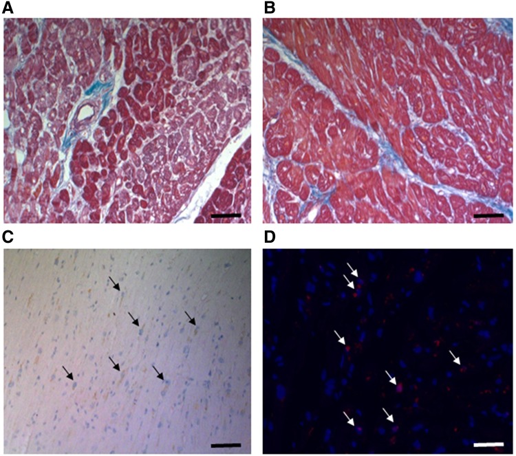
Fig. 1.
Tissue remodeling, apoptosis and oxidative stress in failing human heart sections. Human heart sections were stained by Mason Trichrome (A & B) to analyze collagen deposition (blue) in muscle sections (red). Apoptotic cells were identified by staining for cleaved caspase 3 (C) as indicated in “Materials and methods”, nuclei were counterstained using hematoxylin. Finally, oxidative damage was visualized by staining for 8oxoG lesions (D) by fluorescence immunohistochemistry (red) with DAPI counterstained nuclei (blue). Black bars represent 100 µm, white bar 50 µm

