Fig. 4.
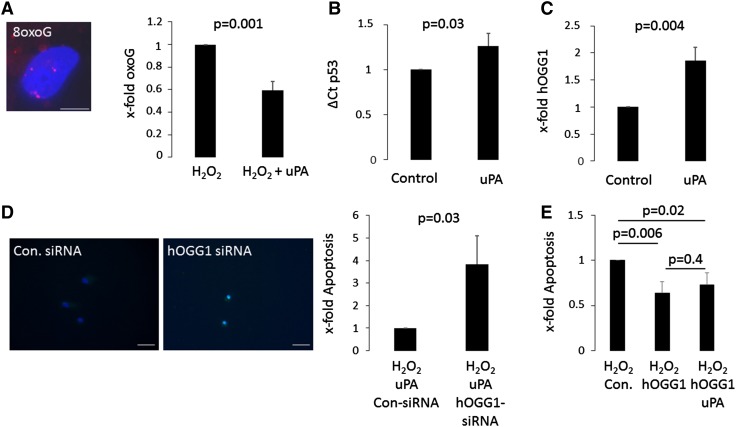
uPA protects human adult cardiac myocytes via hOGG1. Human adult cardiac myocytes (HACM), pretreated with 100 U/ml uPA for 24 h or left untreated, were incubated with 200 µM H2O2 for 2 h and 8-oxoguanine (8oxoG) lesions in cellular nuclei were determined after 24 h by immunofluorescence staining as indicated in “Materials and methods”. Values are given as x-fold oxoG lesions and represent mean of 3 determinations ± SD. The inset shows a representative image of a nucleus with 8oxoG lesions in red (A, bar represents 20 µm). mRNA levels for p53 were determined by qPCR after 2 h and normalized to GAPDH (B) and levels for hOGG1 (C) were determined by flow cytometry after 24 h of uPA treatment as indicated in “Materials and methods”. Values are given as ΔCt for p53 (B) and x-fold mean fluorescent intensity of hOGG1 (C) over untreated controls and represent mean of 3 determinations ± SD. HACM, transfected with hOGG1-siRNA or with control-siRNA as described in “Materials and methods” and pretreated with 100 U/ml uPA for 24 h were incubated with 200 µM H2O2 for 2 h. Apoptosis was quantified as described in “Materials and methods”. Values are given as x-fold apoptotic cells over control and represent mean of 3 determinations ± SD. The inset shows representative TUNEL staining images of the different conditions, the white bar represents 60 µm (D). HACM overexpressing hOGG1 or a control vector (Con.) were incubated with 200 µM H2O2 for 2 h. Apoptosis was quantified as described in “Materials and methods”. Values are given as x-fold apoptotic cells over control and represent mean of 3 determinations ± SD (E)

