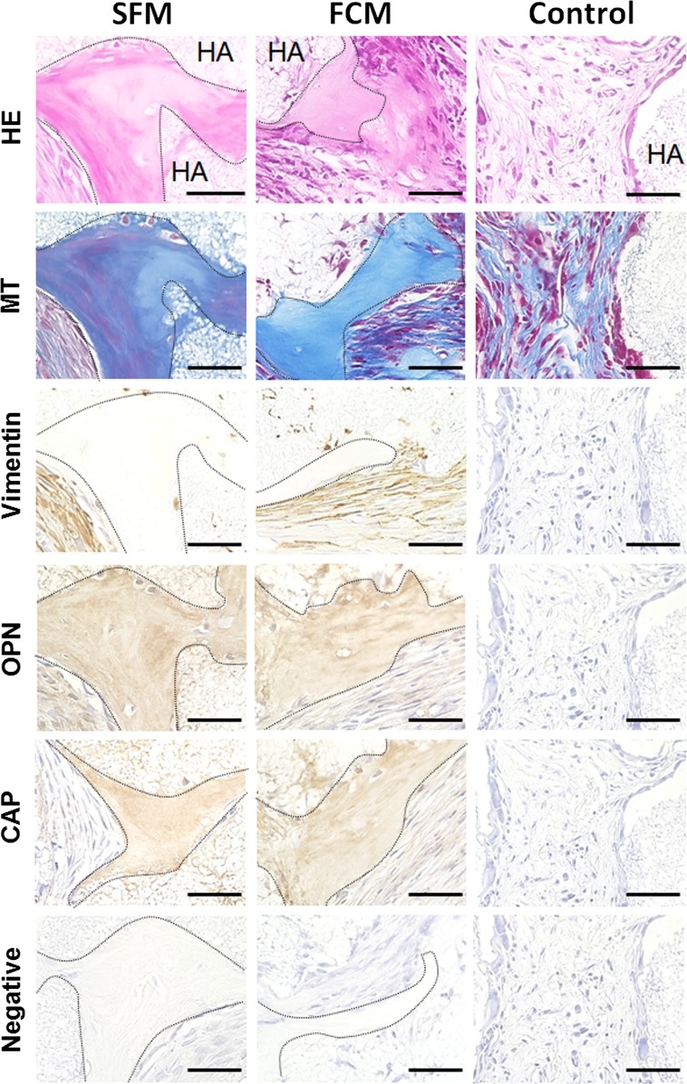
Fig. 4.
In vivo transplantation of ex vivo-expanded PDLSCs using serum-free- or FBS-containing medium; histological and immunohistochemical evaluation of cell/hydroxyapatite (HA)-scaffold constructs containing PDLSCs cultured in serum-free (SFM) or FBS-containing (FCM) medium in nude mice after subcutaneous transplantation. Hematoxylin and eosin (HE) and Masson’s trichrome (MT) staining indicated that the newly formed cementum hard tissue, which is indicated by the dashed line, was observed at the surface of HA-scaffold carrier (HA) in the SFM- and FCM-cell constructs. Immunoreactivity for the human-specific vimentin antibody was detected in the fibroblasts in connective tissue around the de novo hard tissue in both types of cell/scaffold constructs. Moreover, the hard tissue matrix was positively immunostained with antibodies against osteopontin (OPN) and cementum attachment protein (CAP), which are typical markers for cementum hard tissue. Neither hard tissue formation nor immunoreactivity for any antibodies was observed in the HA-scaffold alone (Control). The primary antibody was omitted during immunostaining (Negative); scale bars 100 µm

