Figure 3.
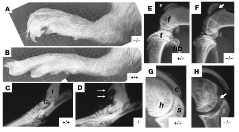
Clinical appearance and radiographic changes in Prg4–/– mice. Photograph of the hind paws of 6-month-old Prg4–/– (A) and wild-type (B) mice. Note the curved digits in the mutant mouse and the swelling at the ankle joint. (C and D) Radiograph of the ankle joint of 9-month-old wild-type and Prg4–/– mice, respectively. The structures corresponding to the tibia (t) and talus (ta) are indicated. Note the calcification of structures adjacent to the ankle (white arrows). (E) Lateral knee x-ray of a 4-month-old wild-type mouse. The structures corresponding to the patella (p), femoral condyle (f), tibial plateau (t), and fibula (fib) are indicated. (F) Lateral knee x-ray of a 4-month-old Prg4–/– mouse. Note the increased joint space between the patella and femur (arrowheads) and osteopenia of the patella, femoral condyles, and tibial plateau. (G) Shoulder x-ray of a 4-month-old wild-type mouse. The structures corresponding to the humeral head (h), glenoid fossa of the scapula (s), and lateral portion of the clavicle (c) are indicated. (H) Shoulder x-ray of a 4-month old Prg4–/– mouse. Note the increased joint space between the humerus and scapula (arrowheads) and the osteopenia of the humeral head.

