Figure 3.
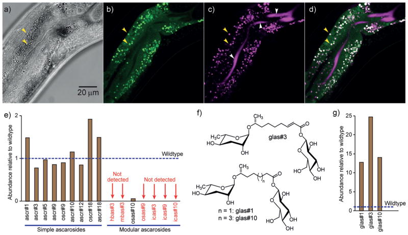
Identification of LROs as the site of the biosynthesis of modular ascarosides. a) A bright-field image of the midsection of a transgenic worm expressing gfp::acs-7 under the acs-7 promoter in acs-7 mutant background (young adult stage). b) GFP expression is localized to punctate organelles (e.g., yellow arrows). c) Staining of LROs with LysoTracker Red (white arrows: background staining of the intestinal lumen). d) Overlay of images in (b) and (c). e) Abundances of ascarosides in glo-1(zu437) mutants relative to wildtype C. elegans. f) Structures of ascaroside glucosyl conjugates. g) Abundances of the glucosyl conjugates glas#1, glas#3, and glas#10 are greatly increased in glo-1 knockout mutants. In (e) and (g), the values shown represent the averages of two replicates.

