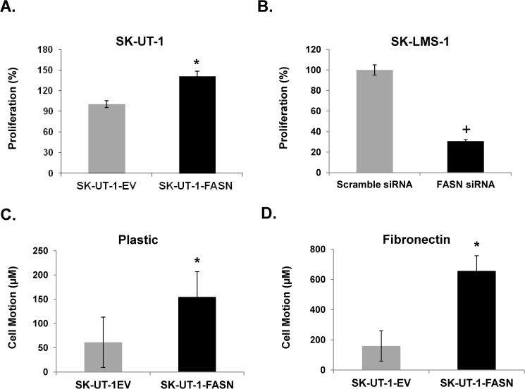
Fig 5. FASN promotes Ut-LMS proliferation and increases Ut-LMS motion.
(A) DIMSCAN assay in SK-UT-1-EV and -FASN cells after 72 hrs incubation in 96 wells plate; (B) DIMSCAN assay in FASN siRNA-transfected SK-LMS-1 cells at 72 hr. Quantified cell motion detected by 72 hr time-lapse video microscopy. (C)Plastic plate; (D)Fibronectin-coated plate. Cell motion was recorded, tracked and quantitated by Image-Pro Premier 9.1.4. *, p<0.05 FASN-transduced SK-UT-1 vs. EV-transduced SK-UT-1 cells; +, p<0.05 FASN siRNA transfected SK-LMS-1 vs. scramble siRN transfected SK-LMS-1 cells.

