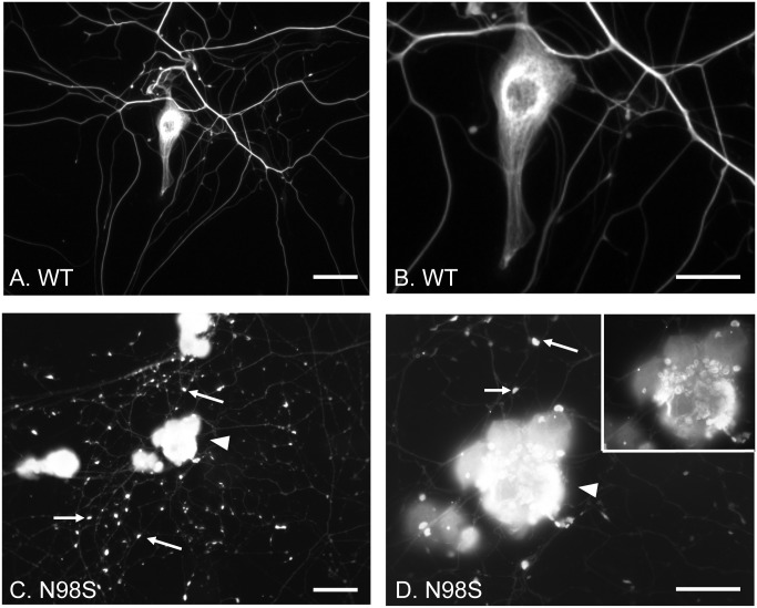
Fig 1. Immunofluorescence micrographs of cultured DRG neurons from Nefl+/+ and NeflN98S/+ mice labeling NFL.
(A and C) Low power views of DRG neurons from Nefl+/+ (A) and NeflN98S/+ (C) mice. Note that in NeflN98S/+ DRG neurons, the processes are characterized by large amounts of enlarged and bright particles along the processes, pointed by arrows (C). (B and D) High-magnification images of DRG neurons as seen in Fig A and C. Note the filamentous structure in Nefl+/+ DRG neuron (B). In contrast, disrupted neurofilament network and enlarged neurofilament particles (pointed by arrows) along the processes can be seen in NeflN98S/+ DRG neurons (D). A low intensity image of DRG neurons pointed by the arrowhead is shown in the inset (D) that also shows the broken neurofilamentous network. NeflN98S/+, n = 8; Nefl+/+, n = 5. Scale bars = 50 μm (A and C) and 25 μm (B and D).

