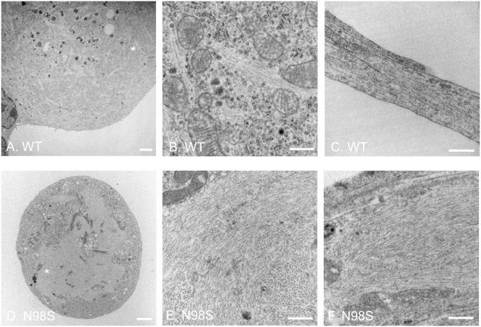
Fig 5. Electron micrographs of cultured DRG neurons from wild-type and NeflN98S/+ mice.
(A and B) EM of cell soma of wild-type. Fig B is a higher magnification view of the asterisk-labeled area in Fig A. Filamentous structures formed by intermediate filaments can been seen throughout the cell body, next to organelles such as mitochondria. (C) A longitudinal section of processes of Nefl+/+ DRG. Bundles of microtubules and intermediate filaments are running in parallel alongside the processes. (D and E) EM of cell soma of NeflN98S/+ DRG. Fig E is a higher magnification view of the asterisk-labeled area in Fig D. Massive accumulation of disordered neurofilaments is observed in the soma of NeflN98S/+ DRG (D). The density of neurofilaments is very high and few other cytoplasmic elements can be seen within the accumulations (E). (F) A longitudinal section of processes of NeflN98S/+ DRG. An enlarged process area of disorganized filamentous accumulation can be seen. Scale bars = 2 μm (A and D) and 500 nm (B, C, E and F).

