. 2017 Jun 27;6:e24139. doi: 10.7554/eLife.24139

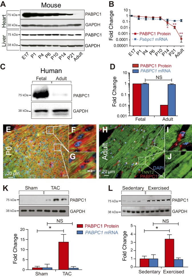
Figure 1. PABPC1 is dynamically regulated during cardiac development and hypertrophy.

(A–D) Relative quantification of PABPC1 protein (immunoblots) and mRNA (qPCR) levels normalized to GAPDH during mouse heart and liver development (A, B) and in human fetal and adult hearts (C, D). (E–J) Immunofluorescent images of mouse postnatal day 0 (P0) and 8-week-old adult hearts stained for PABPC1 (red), cardiac troponinT (green), and DAPI (blue). Insets G and J show cardiomyocytes, while F and I show non-cardiomyocytes. Immunoblots and quantification of PABPC1 protein and mRNA from wild-type mouse hearts 8 weeks after (K) thoracic aortic constriction (TAC) or (L) 10-week exercise training. Data are mean ± s.d (n = 3); *p<0.05, unpaired two-tailed t-test. NS, not significant.

DOI: http://dx.doi.org/10.7554/eLife.24139.003

Figure 1.

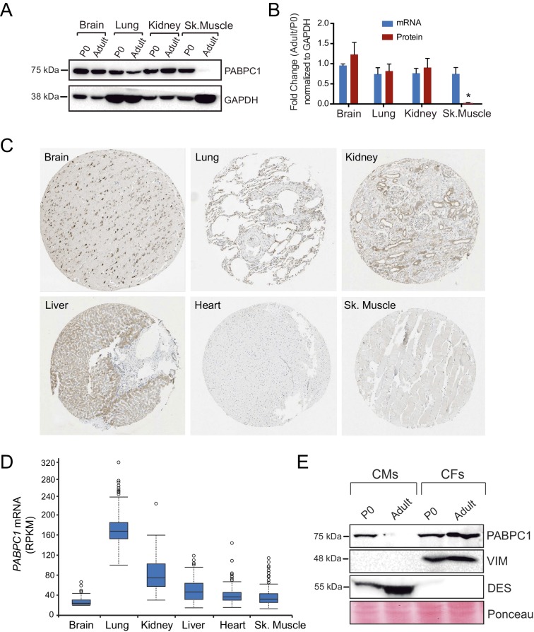
Figure 1—figure supplement 1. Post-transcriptional silencing of PABPC1 is muscle-specific.

Figure 1—figure supplement 1.

(A) Immunoblots for PABPC1 from postnatal day 0 (P0) and 8-week-old adult mice from the indicated tissue with GAPDH as a loading control. (B) Quantification of PABPC1 protein and mRNA expression (qPCR). Data are mean ± s.d (n = 3); *p<0.005 unpaired two-tailed t-test. (C) PABPC1 immuno-histochemistry images based on anti-PABPC1 antibody (Abcam ab21060) from the Human Protein Atlas Database (www.proteinatlas.org) showing that PABPC1 is abundantly expressed in other adult tissues but not heart and skeletal muscle. (D) RPKM values from the Human Protein Atlas Database showing relative Pabpc1 mRNA levels in various human tissues. (E) Western blot of purified P0 and adult cardiomyocytes (CMs) and cardiac fibroblasts (CFs). Blotting for Desmin (DES, CM marker) and Vimentin (VIM, CF marker) show clean separation of cell types.

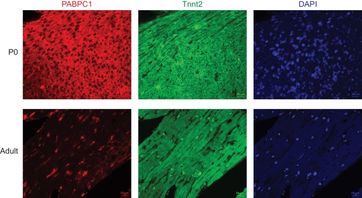
Figure 1—figure supplement 2. Post-transcriptional silencing of PABPC1 is muscle-specific.

Figure 1—figure supplement 2.

Single-channel immunofluorescent images of mouse postnatal day 0 (P0) and 8-week-old adult hearts stained for PABPC1 (red), cardiac troponinT (green), and DAPI (blue).