
Figure 3.
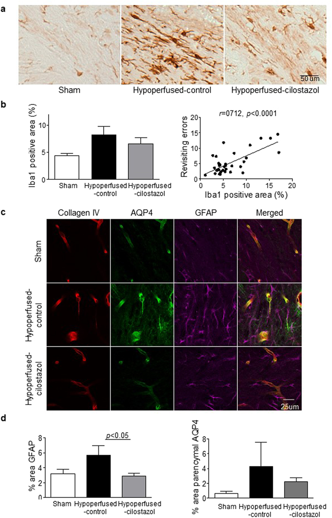
Cilostazol reduced the extent of gliosis. (a) Representative images of Iba1 staining in the corpus callosum. (b) There was a trend towards an increase in Iba1 stained micoglia that was reduced with cilostazol after hypoperfusion. There was a robust association between microgliosis and the number of revisiting errors (Pearson r = 0.712, p < 0.0001). (c) Representative images of immunofluorescence for Collagen IV, AQP4 and GFAP in the corpus callosum. (d) There was an increase in the area of GFAP positive astroglia in the hypoperfused-control mice. There was a significant reduction with cilostazol treatment as compared to control hypoperfused. Astrocyte-endfoot displacement tended to be observed in the hypoperfused-control mice and less in the cilostazol treated mice. Data represents mean ± SEM. *p < 0.05, Hypoperfused-control vs Hypoperfused-cilostazol.