
Figure 4.
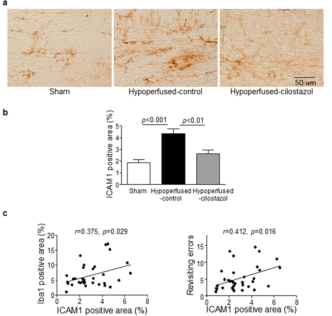
Cilostazol significantly suppressed endothelial adhesion molecule expression in the corpus callosum. (a) Representative images of intercellular adhesion molecule-1 (ICAM1) staining in the corpus callosum. (b) There was a significant difference in ICAM staining between the three groups (F (2, 33) = 13.19; p < 0.0001). ICAM1 immunostaining was significantly greater in the hypoperfused-control group compared to the sham (p < 0.001) and this was reduced with cilostazol treatment (p < 0.01). (c) There was a significant association between ICAM1 positive areas and microgliosis (Pearson r = 0.375, p < 0.05), and the number of revisiting errors (Pearson r = 0.412, p < 0.05). Data represents mean ± SEM.