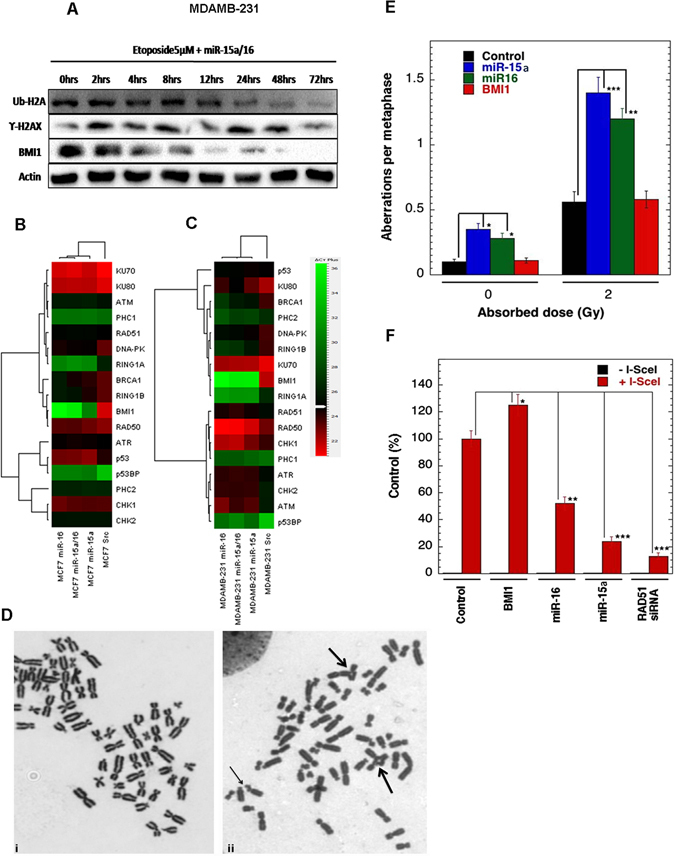
Figure 5.

Time-dependent expression of proteins after ectopic expression of miR-15a/16 and overexpression of miR-15a/16 impedes homologous recombination-mediated repair. Western blot analysis showing the expression of BMI1, γ-H2AX, Ub-H2A in MDAMB-231 cells transfected with both miR-15a/16 followed by treatment with etoposide for 2 to 72 hrs. Actin was used as a gel loading control. During the course of time periods (0 hrs to 72 hrs). Blots were cropped to enhance the representation (A). Heat map showing the endogenous expression of quantitative RT-PCR analysis. The Green colour in the heat map indicates decreased and red colour indicates increased mRNA expression level. Gene specific primers were used for the amplification. GAPDH was used as endogenous control (B,C) Metaphase chromosomes of cells overexpressing Mir-15a/16. (i) Metaphase fromcontrol, (ii) Metaphase from irradiated cell showing gaps (small arrow) and radials (large arrows)in human H1299 cells (D). In human H1299 cells S-phase specific chromosome aberrations after 2 Gy irradiations. *p < 0.05, **p < 0.01, ***p < 0.001 Student t-test (E) Homologous recombination (HR) repair of DNA DSB repair in human H1299 cells are shown with and without I-SceI induction.*p < 0.05, **p < 0.01, ***p < 0.001Student t-test (F).
