
Figure 6.
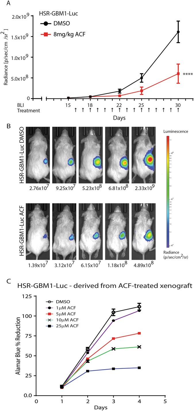
ACF inhibits the growth of small, visually undetectable, HSR-GBM1-Luc xenograft growth in vivo. (A) HSR-GBM1-Luc xenografts were grown for 15 days. Treatment was administered by intraperitoneal injections of vehicle (DMSO in saline) or ACF (8 mg/kg) for 15 injections (indicated by arrows in (A)). Averaged luciferase activity (radiance) is shown. ****p < 0.0001 ACF vs. vehicle (two-way ANOVA with Sidak’s multiple comparisons test). (B) BLI of representative (of the mean) animals are shown for the vehicle (top) and ACF-treated animals (bottom). (C) Alamar Blue in vitro growth assay was performed on HSR-GBM1-Luc cells freshly isolated from ACF-treated animals. Cells were treated in hypoxia (1% oxygen) with 0 (DMSO), 1, 5, 10, and 25 μM ACF. A representative analysis is shown.