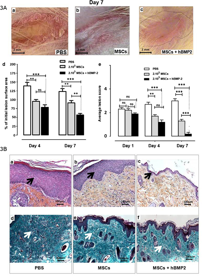
Figure 3.

Effect of hBMP-2 addition to MSC therapy on radiation-induced dermatitis repair. (A) Photographs of radiation-induced lesions 7 days after injection. Three groups of animals were irradiated at 55 Gy and received PBS (PBS), MSCs (MSCs), or MSCs and hBMP-2 (MSCs + hBMP-2) infusion three weeks later. (a) PBS treatment, (b) infusion of 2 × 10⁵ MSCs, and (c) co-injection of 2 × 10⁵ MSCs and hBMP-2. (d) % of initial lesion surface area and (e) average lesion scores. PBS (white bars), MSCs (grey bars), or MSCs + hBMP-2 (black bars). (B) One week after treatment, HES staining (a–c) (a) PBS control infusion; (b) local infusion of 2 × 10⁵ MSCs; and (c) hBMP-2 co-infusion with MSCs. (d–f) Masson’s Trichrome staining of: (d) PBS control infusion; (e) local infusion of 2 × 10⁵ MSCs; and (f) hBMP-2 co-infusion with MSCs. Each group of animals comprised 10 Sprague Dawley rats. Comparison by ANOVA followed by the Mann-Whitney or Dunnett’s test for group pair-wise comparisons. Data represent mean ± SEM, ns: not significant, *p < 0.05, **p < 0.01, ***p < 0.001.
